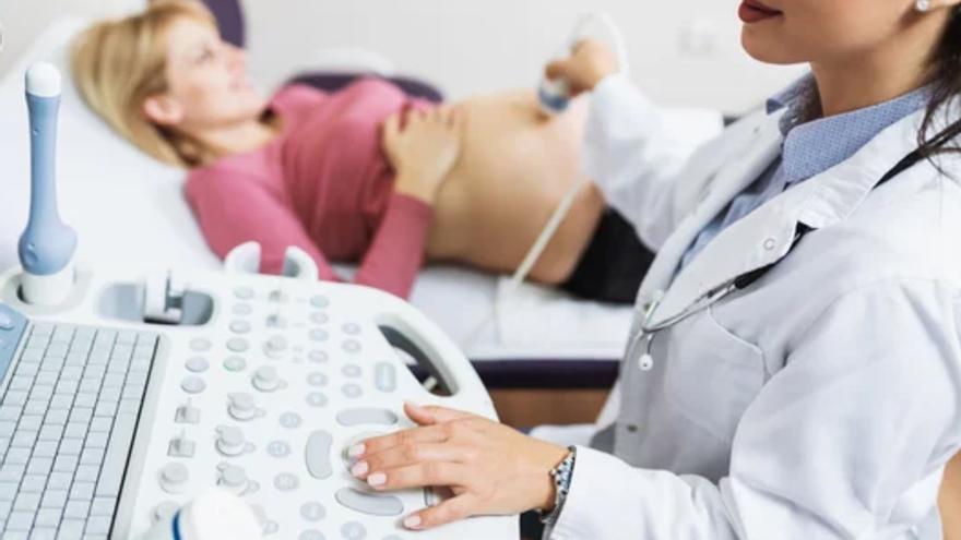
Pregnancy or prenatal control It is medical monitoring that is carried out throughout the pregnancy period with the aim of: Preventing diseases or diagnosing them early. Thus, the health of the fetus and mother is protected, and this is achieved Natural development From pregnancy today, gynecologists have made progress in this field, e.g Genetic studies And the AI-powered ultrasonic control.
the doctor Fermin Esteban Navarrogynecologist and specialist in prenatal diagnosis and fetal medicineRoper International Hospital“, explains what are the levels of prevention for correct pregnancy control: “Primary prevention that is carried out through Pre-pregnancy advice; Secondary prevention based on Conducting tests and examination Which allows us to evaluate the risks of various problems between the mother and the fetus; The third prevention is trying to start a Early treatment of the identified problem “As quickly as possible in order to minimize the damage caused by the disease.”
Preventing preeclampsia
Example Secondary prevention It is the examination, or Investigatel revealing of High risk of preeclampsia. Preeclampsia is a pregnancy complication that can become serious if it develops EclampsiaWhich puts the lives of the mother and fetus at risk. The main symptoms are Arterial hypertensionAccompanied by protein in the urine.
Screening for preeclampsia consists of a Doppler echocardiography From the maternal uterine arteries, which take place in The twelfth week Of pregnancy during ultrasound, explains Dr. Esteban Navarro The shape of the fetus and the baby's heartAnd other possible tests, such as chromosomal diseases.
The doctor explains that the test, along with studying other risk factors such as age, “allows us to detect pregnant women who are at high risk of developing preeclampsia with a sensitivity of up to 70%. This helps us give Preventive treatments And guide the patient as best we can Prevent the development of the disease“If there is suspicion of preeclampsia, complementary tests are performed, such as determining vascular factors, which help in the diagnosis and measure the level of risk of possible complications.
Is it possible to detect premature birth?
Although this is true The premature part o Prematurity is not a disease, but it can be one Endangering the life of the fetusAnd in some circumstances Hello from the sea. Obviously, the longer the birth, the greater the risks to the baby. The answer to the question is yes: yes can be detected.
Dr. Esteban Navarro explains that the assessment of the risk of premature birth has been done so far, “by measuring… Cervical length Through a Ultrasound In which we perform Week 20 Of pregnancy if the neck is less than that 25 mm We consider that there is Other risks “From premature birth, if it measures more than 35 mm, we consider the risk to be low.”
The doctor adds that they now have ultrasound tools, such as the test Early quantitiesis used to estimate Cervical tissue Thus assessing the risk of premature birth. This image evaluation technology is supported by Artificial intelligence algorithms. The test is performed between weeks 19 and 24 of pregnancy.
What genetic tests are used during pregnancy?
AndGenetic testingIt is an innovative preventive tool that allows you to know whether a person is infected or not Genetic predisposition to contracting a disease, or provide some type of characteristics that help in early diagnosis of pathology. It allows, for example, to obtain information about Intolerance Food, prevent Diabetes Or even a diagnosis of alopecia.
Within load range you can do this Prenatal testing Non-invasively in order to evaluate the fetus's risk of aneuploidy, ie. Numerical chromosomal changes, on chromosomes 13, 18 and 21 or on the X and Y sex chromosomes. There are more complex tests that also evaluate the risk of having another type of change in all chromosomes. You only need one to run the tests Blood sample From the beach.
Emotional ultrasound: 3D and 4D ultrasound
Since the world around us has in principle only three dimensions, it is interesting to take a few seconds to understand it What is it 4D ultrasound. We start from the classic 2D ultrasound, in black and white and in 2D, which is still very important to evaluate the correct conformation, growth and development of the baby, and is performed in real time.
3D and 4D ultrasound are performed with the same sonographer. 3D They brought us the third dimension Depthin a still image, and 4D adds real-time motion.
the doctor Miguel Angel LopezTeam coordinator Obstetrics and Gynecology at Quirónsalud Toledo HospitalExplains the advantages of ultrasound performed with highly accurate and equipped equipment Doppler and 3D-4D technology. 3D allows structures to be visualized with great precision. If four dimensions are added, it is possible to identify many fetal movements, of the head, face, and limbs.
This type of technology is also called… Emotional ultrasoundBecause it helps tighten Emotional bond Between parents and future child. Thanks to 3D-4D technology, Dr. Lopez explains, “another interesting aspect emerges in the study Facial expressions It is a valuable tool for detecting anatomical diseases with a unique ability to explore and identify appropriate neurophysiological development of the fetus, and thus it has a tremendous amount of potential. Potential in perinatal research“.
If what you are looking for when doing 3D and 4D ultrasound is just to show the baby to the future parents, the best images will be obtained between Week 25 and week 30. It is important to emphasize that 3D and 4D ultrasound are complementary to the basic tests in any pregnancy.
What is 5D ultrasound?
5D ultrasound Add to all of the above, Game of lights and shadowsWhich allows for estimation Textures and colors And get a more specific and more accurate picture of the child. At Hospital Quirónsalud Málaga, they have 5D ultrasound, to which they added 5G technology last year.
According to the doctor Rodrigo Orozco Fernandezhead of the obstetrics and gynecology service at the center in Malaga: “5D ultrasound allows you to visualize The fetus in size and movementBut the ultimate disruptor and innovation is the integration of 5G into pregnancy ultrasound. In this way, power becomes possible Share in real time Broadcast your ultrasound with family and friends around the world.” 5D ultrasound with 5G technology Latency from the time elapsed between what is watched and what is streamed is minimized.
Dr. Orozco Fernandez comments that at the Malaga Center they perform regular ultrasound at every consultation and high-resolution ultrasound at Weeks 12, 20 and 34. In addition, it offers the possibility of high-resolution 5D and 5G ultrasound imaging between Week 28 and 32The stage of pregnancy where there is a better view of the fetus.
The advantage of 5D ultrasound is that it is shown from one sideThe real appearance of the child. But it also works to create Morphological study of all organs and systems. It is a complementary test, with diagnostic value, for Ultrasound protocol for pregnancy monitoring.

“Infuriatingly humble social media buff. Twitter advocate. Writer. Internet nerd.”


