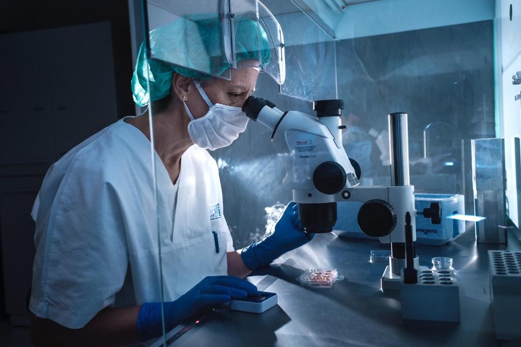
Published in the May issue of the journal Nature Protocols, part of the journal Associated with the prestigious journal Nature, research findings demonstrate the experimental protocol for obtaining in vitro three-dimensional structures, called organoids, that resemble mini-brains capable of developing optic vesicles, the structures that precede the formation of eye, through a process similar to what occurs during embryonic development.
The study, in which researchers Giuliano Callini and Maria Giovanna Reparbelli of the Department of Life Sciences of the University of Siena participated, was headed by Guy Gopalakrishnan of Heinrich Heine University Dusseldorf (Germany).
Organelles aren’t true organs, but tiny structures, tiny groups of cells that scientists consider medicine’s new frontier. These 3D structures represent very promising study models in scientific research because although they are less complex than the original organ, they are probably more representative than conventional cell culture.
Studying organogenesis can help understand the mechanisms that underlie the development of different organs and can provide valuable insights into how these organs interact with specific drugs or therapies. Organoids can be used to identify new biomarkers, screen new drugs, and determine the best future treatment for a patient, with the goal of increasingly precise and personalized medicine.
The experimental method developed by the researchers represents a significant improvement in this sector, as to date most protocols have allowed brain or retinal organoids to be obtained separately.
In this article, the authors of the paper induced neural differentiation in induced pluripotent stem cells that acquire neural envelopes and which, under certain experimental conditions, organize themselves to form brain organelles that subsequently develop optic vesicles.
The development of the protocol required the collaboration of knowledge and skills of various kinds which allowed this important result to be achieved. The Research Unit of the Department of Life Sciences has conducted ultrastructural studies of photovesicle-cerebral organoids, using both transmission electron and scanning microscopy, allowing an understanding of the precise organization of the organoid and the identification of the different cellular classes that make it up.

“Infuriatingly humble social media buff. Twitter advocate. Writer. Internet nerd.”


